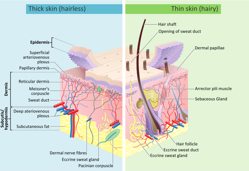
Cross-section
Development
Skin colour
Main article: Human skin color
Human skin shows high skin color variety from the darkest brown to the lightest pinkish-white hues. Human skin shows higher variation in color than any other single mammalian species and is the result of natural selection. Skin pigmentation in humans evolved to primarily regulate the amount of ultraviolet radiation (UVR) penetrating the skin, controlling its biochemical effects.
The actual skin color of different humans is affected by many substances, although the single most important substance determining human skin color is the pigment melanin. Melanin is produced within the skin in cells called melanocytes and it is the main determinant of the skin color of darker-skinned humans. The skin color of people with light skin is determined mainly by the bluish-white connective tissue under the dermis and by the hemoglobin circulating in the veins of the dermis. The red color underlying the skin becomes more visible, especially in the face, when, as consequence of physical exerciseor the stimulation of the nervous system (anger, fear), arterioles dilate.
There are at least five different pigments that determine the color of the skin. These pigments are present at different levels and places.
- Melanin: It is brown in color and present in the basal layer of the epidermis.
- Melanoid: It resembles melanin but is present diffusely throughout the epidermis.
- Carotene: This pigment is yellow to orange in color. It is present in the stratum corneum and fat cells of dermis and superficial fascia.
- Hemoglobin (also spelled haemoglobin): It is found in blood and is not a pigment of the skin but develops a purple color.
- Oxyhemoglobin: It is also found in blood and is not a pigment of the skin. It develops a red color.
There is a correlation between the geographic distribution of UV radiation (UVR) and the distribution of indigenous skin pigmentation around the world. Areas that highlight higher amounts of UVR reflect darker-skinned populations, generally located nearer towards the equator. Areas that are far from the tropics and closer to the poles have lower concentration of UVR, which is reflected in lighter-skinned populations.
In the same population it has been observed that adult human females are considerably lighter in skin pigmentation thanmales. Females need more calcium during pregnancy and lactation and vitamin D which is synthesized from sunlight helps in absorbing calcium. For this reason it is thought that females may have evolved to have lighter skin in order to help their bodies absorb more calcium.
The Fitzpatrick scale is a numerical classification schema for human skin color developed in 1975 as a way to classify the typical response of different types of skin to ultraviolet (UV) light:
| I | Always burns, never tans | Pale, Fair, Freckles |
| II | Usually burns, sometimes tans | Fair |
| III | May burn, usually tans | Light Brown |
| IV | Rarely burns, always tans | Olive brown |
| V | Moderate constitutional pigmentation | Brown |
| VI | Marked constitutional pigmentation | Black |
Aging
For more details on this topic, see senescence.
For more details on this topic, see Intrinsic and extrinsic aging.
As skin ages, it becomes thinner and more easily damaged. Intensifying this effect is the decreasing ability of skin to heal itself as a person ages.
Among other things, skin aging is noted by a decrease in volume and elasticity. There are many internal and external causes to skin aging. For example, aging skin receives less blood flow and lower glandular activity.
A validated comprehensive grading scale has categorized the clinical findings of skin aging as laxity (sagging), rhytids (wrinkles), and the various facets of photoaging, including erythema (redness), and telangiectasia, dyspigmentation (brown discoloration), solar elastosis (yellowing), keratoses (abnormal growths) and poor texture.
Cortisol causes degradation of collagen, accelerating skin aging.
Anti-aging supplements are used to treat skin aging.
Photoaging
Main article: Photoaging
Photoaging has two main concerns: an increased risk for skin cancer and the appearance of damaged skin. In younger skin, sun damage will heal faster since the cells in the epidermis have a faster turnover rate, while in the older population the skin becomes thinner and the epidermis turnover rate for cell repair is lower which may result in the dermis layer being damaged.
Functions
Skin performs the following functions:
- Protection: an anatomical barrier from pathogens and damage between the internal and external environment in bodily defense; Langerhans cells in the skin are part of the adaptive immune system.
- Sensation: contains a variety of nerve endings that react to heat and cold, touch, pressure, vibration, and tissue injury; see somatosensory system and haptics.
- Heat regulation: the skin contains a blood supply far greater than its requirements which allows precise control of energy loss by radiation, convection and conduction. Dilated blood vessels increase perfusion and heatloss, while constricted vessels greatly reduce cutaneous blood flow and conserve heat.
- Control of evaporation: the skin provides a relatively dry and semi-impermeable barrier to fluid loss. Loss of this function contributes to the massive fluid loss in burns.
- Aesthetics and communication: others see our skin and can assess our mood, physical state and attractiveness.
- Storage and synthesis: acts as a storage center for lipids and water, as well as a means of synthesis of vitamin D by action of UV on certain parts of the skin.
- Excretion: sweat contains urea, however its concentration is 1/130th that of urine, hence excretion by sweating is at most a secondary function to temperature regulation.
- Absorption: the cells comprising the outermost 0.25–0.40 mm of the skin are "almost exclusively supplied by external oxygen", although the "contribution to total respiration is negligible". In addition, medicine can be administered through the skin, by ointments or by means of adhesive patch, such as the nicotine patch oriontophoresis. The skin is an important site of transport in many other organisms.
- Water resistance: The skin acts as a water resistant barrier so essential nutrients aren't washed out of the body.
Skin flora
Main article: Skin flora
The human skin is a rich environment for microbes. Around 1000 species of bacteria from 19 bacterial phyla have been found. Most come from only four phyla: Actinobacteria (51.8%), Firmicutes (24.4%), Proteobacteria (16.5%), andBacteroidetes (6.3%). Propionibacteria and Staphylococci species were the main species in sebaceous areas. There are three main ecological areas: moist, dry and sebaceous. In moist places on the body Corynebacteria together withStaphylococci dominate. In dry areas, there is a mixture of species but dominated by b-Proteobacteria andFlavobacteriales. Ecologically, sebaceous areas had greater species richness than moist and dry ones. The areas with least similarity between people in species were the spaces between fingers, the spaces between toes, axillae, and umbilical cord stump. Most similarly were beside the nostril, nares (inside the nostril), and on the back.
Reflecting upon the diversity of the human skin researchers on the human skin microbiome have observed: "hairy, moist underarms lie a short distance from smooth dry forearms, but these two niches are likely as ecologically dissimilar as rainforests are to deserts."
The NIH has launched the Human Microbiome Project to characterize the human microbiota which includes that on the skin and the role of this microbiome in health and disease.
Microorganisms like Staphylococcus epidermidis colonize the skin surface. The density of skin flora depends on region of the skin. The disinfected skin surface gets recolonized from bacteria residing in the deeper areas of the hair follicle, gut and urogenital openings.


0 comments:
Post a Comment